When the veins in our legs become prominent from the outside, ache, or swell, the first solution that usually comes to mind is to eliminate that superficial appearance. However, modern vascular surgery has taught us that visible varicose veins are actually just the tip of the iceberg. The real problem often lies deep beneath the skin, in invisible valve insufficiencies or the backward leakage of blood (reflux). At this precise point, Doppler Ultrasound, which serves as the “compass” of varicose vein treatment, comes into play.
Op. Dr. Nebiye Tüfekçi Varer, with her expertise in cardiovascular surgery at her clinic in Kartal, maps out the veins by performing a detailed Doppler examination for every patient before starting treatment. This is because a treatment started without a correct diagnosis offers only a temporary solution; permanent recovery is only possible by eliminating the root cause of the problem.
What is Doppler Ultrasound and How Does It Work?
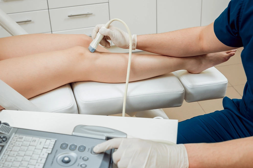
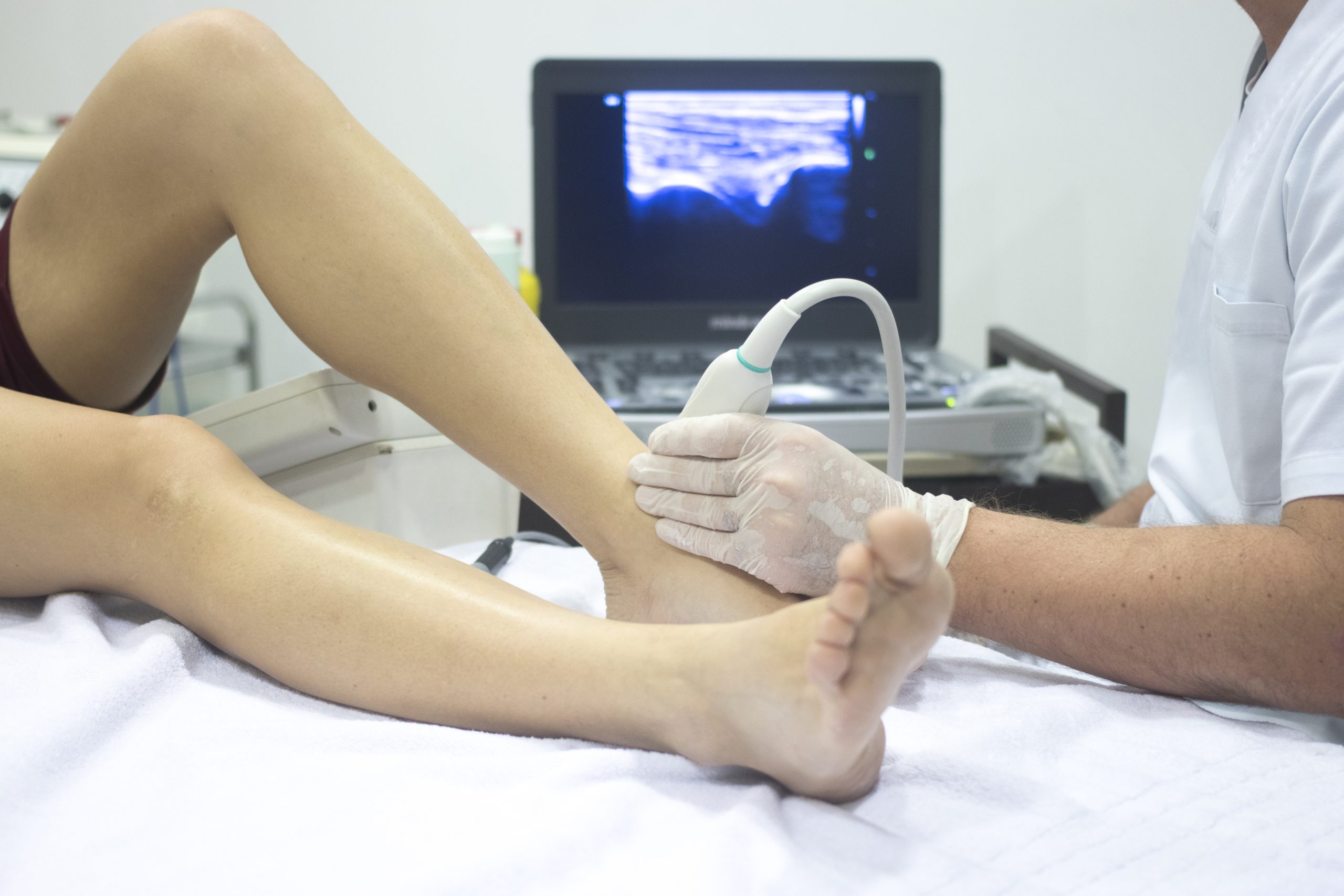
Doppler ultrasound is a technology that allows us to monitor blood flow within the veins both visually and audibly, using the power of sound waves. Thanks to this advanced imaging method used nowadays, the internal structure and width of the veins, and most importantly, the direction of blood flow, can be monitored in real-time.
-
Color-Coded Flow: The red and blue colors you see on the screen tell us whether the blood is moving toward the heart or succumbing to gravity and leaking back toward the legs.
-
Audible Feedback: The speed and rhythm of the flow within the vein are tracked by sound, analyzing how healthily the valves are functioning.
Why is it Indispensable in Varicose Vein Treatment?
Many people might think, “I only have spider veins, do I really need an ultrasound?” However, we know today that a small spider vein on the surface can be caused by insufficiency in a large deep vein.
-
Finding the Source of the Problem: Varicose veins occur when the valves inside the veins malfunction. Doppler ultrasound identifies exactly at what level this leakage (reflux) begins without error.
-
Personalized Treatment Plan: Every leg structure and every vascular issue is unique. Thanks to ultrasound data, whether laser, sclerotherapy, or foam treatment will be applied to you is decided with surgical precision.
-
Preventing Risks: Vital risks that are not visible to the naked eye, such as deep vein thrombosis (clots), can only be diagnosed early through a Doppler examination.
Kartal Varicose Vein Treatment: Imaging Through a Surgeon’s Eyes
The ultrasound device is a tool, but interpreting the data on that screen requires true expertise. Concentrating on vascular health in the Kartal region, Op. Dr. Nebiye Tüfekçi Varer evaluates vascular anatomy with the meticulousness of a surgeon during the Doppler examinations she performs personally.
An accurate varicose vein diagnosis affects the success of the treatment 100%. If needle treatment (sclerotherapy) is applied only to the surface capillaries without detecting the leakage in the main vein, it is inevitable that the varicose veins will recur in a short time. This is why a professional vascular surgeon nowadays uses ultrasound as their “eyes” to plan every step of the treatment based on this data.
What to Expect During the Procedure?
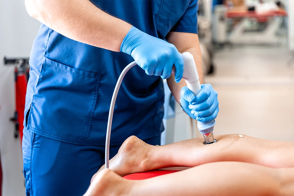
Doppler ultrasound is a completely painless, radiation-free procedure with no side effects. While the tip of the device is moved across your skin with a gel applied to your leg, you can watch the functioning of your own veins on the screen. After this procedure, which takes about 15–20 minutes, a “vascular health report card” for your legs is generated.
If you have swelling, night cramps, a feeling of heaviness, or visible veins in your legs, the healthiest step is to proceed with a scientific diagnosis rather than wasting time with hearsay information. To meet the expertise of Op. Dr. Nebiye Tüfekçi Varer and invest in the future of your legs, you can reach us via nebiyetufekci.com.
The right diagnosis is the beginning of healthy steps.

