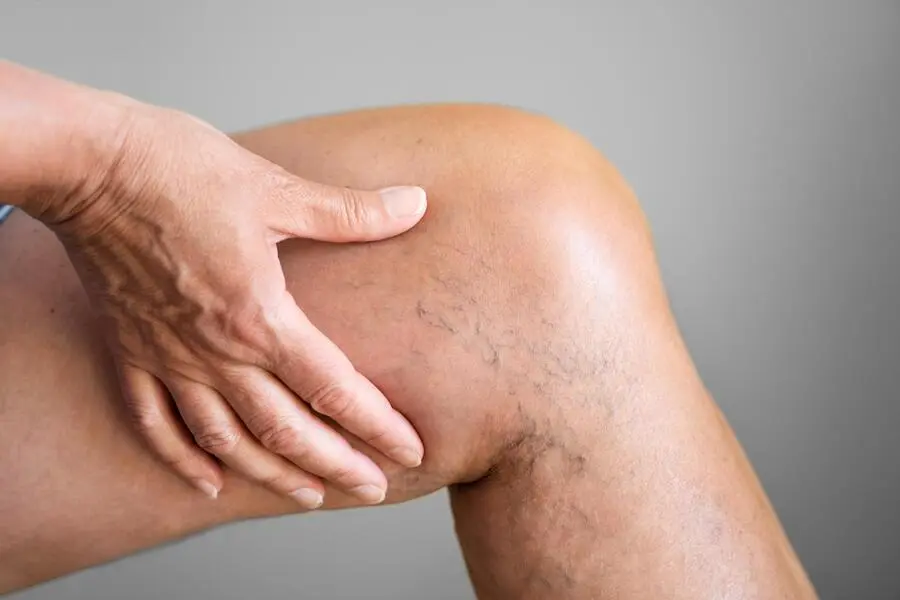
You may be experiencing an increasing feeling of heaviness in your legs later in the day, cramps that interrupt your sleep at night, swelling in your ankles (edema), or purple, green, and red vein networks that are becoming increasingly prominent on the skin surface. Most of the time, our patients try to brush off these complaints as an aesthetic problem or the fatigue of the day. However, varicose veins, that is, venous insufficiency, is a progressive and structural circulatory disease that should be taken seriously.
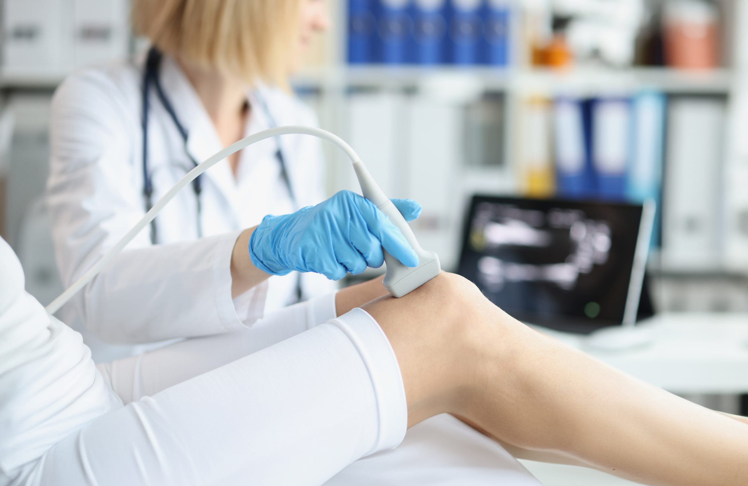
It is impossible to understand from the outside with the naked eye whether this complex circulatory network in our legs is working healthily. The enlarged veins we see on the skin surface are actually just the visible tip of the iceberg. The real problem is the deterioration of the valve system in the main veins that extend deep into the skin and are responsible for carrying blood to the heart. The most fundamental and critical diagnostic tool that makes this invisible world visible to us and allows us to determine the root cause of the disease is the Venous Color Doppler Ultrasonography device.
The most fundamental principle I have adopted in my clinic as a Cardiovascular Surgeon is this: The treatment of no disease that has not been diagnosed correctly can be permanent. In this comprehensive guide, we discuss in detail what vein examination with Venous Color Doppler Ultrasound, the golden standard of varicose vein treatment, is, why it is of vital importance, and how a flawless treatment planning is made.
What is Venous Color Doppler Ultrasound and What Does It Do?
To better understand this concept, it will be useful to break the words into pieces:
- Venous: It refers to the vein system that carries the dirty blood (oxygen depleted blood) in our body from bottom to top, defying gravity, towards the heart.
- Ultrasonography: It is a reliable technology that does not contain radiation, which visualizes the internal structure of tissues using high frequency sound waves that the human ear cannot hear.
- Color Doppler: It works on the principle that sound waves hit the moving blood cells (red blood cells) in the vessel and reflect back. This technology reflects not only the presence of blood but also its flow direction, speed, and character on the screen with red and blue colors.
So What Does It Do? Inside the veins in our legs, there are one way valves that prevent blood from escaping back down (towards the feet) and only open upwards (towards the heart). Over the years, these valves can deteriorate due to genetic predisposition, standing still, pregnancy, or weight gain. When the valves cannot close completely, blood leaks downwards under the influence of gravity and pools in the legs. In medicine, we call this condition “Venous Reflux” (Vein Insufficiency).
Vein Examination with Color Doppler Ultrasound helps us find the answers to the following critical questions:
- Is There Reflux (Leakage)? It measures which vein has valve insufficiency and how long the blood leaks backwards in milliseconds.
- What is the Condition of Vein Diameters? The diameter of a healthy vein and the diameter of a vein that has developed insufficiency and expanded under pressure are different. These diameter measurements determine the treatment method to be applied (Laser, Radiofrequency, Foam, etc.).
- Deep Vein Thrombosis (DVT) Risk: It definitively shows whether there is an old or new clot carrying a vital risk in the deep veins passing through the inner parts of the leg.
How Should a Proper Venous Doppler Ultrasound Be Performed?
We have many patients coming to our clinic with ultrasound reports taken at other centers. However, not every ultrasound examination is sufficient to plan varicose vein treatment. The golden rules of a proper Venous Doppler Ultrasound examination performed with a surgical vision are as follows:
- It Must Absolutely Be Performed Standing: Varicose vein is a “pressure” disease directly related to gravity. When the patient lies on the stretcher, the pressure in the legs instantly drops, the veins shrink, and the leak (reflux) in the valves, if any, is hidden. For this reason, the ultrasound for the diagnosis of venous insufficiency must absolutely be performed while the patient is standing. An ultrasound performed lying down is misleading for the diagnosis of varicose veins.
- Maneuver Tests (Provocation): During the examination, your physician may ask you to bear down (Valsalva maneuver) or squeeze your calf muscles. The physician also applies rhythmic pressure to your calf with their hand. The purpose of these maneuvers is to artificially increase the pressure inside the vein and test how much the valves can resist this pressure, that is, the severity of the leak.
- Connections of Superficial and Deep Systems: There are bridge veins (Perforator Veins) in our legs that connect the superficial veins with the deep veins. Insufficiencies in these bridges should be mapped in detail.
Frequently Asked Questions (FAQ) About Venous Color Doppler Ultrasound
We answer the most frequently encountered questions in order to eliminate the concerns in our patients’ minds regarding the diagnostic process and to make the medical processes transparent:
- Is Doppler Ultrasound imaging a painful or aching procedure? Absolutely not. It is an imaging method performed entirely over the skin by applying a special water based ultrasound gel, without needles or incisions. It does not create any pain, ache, or burning sensation.
- Is there radiation (X Ray) in the procedure? Can pregnant women have it done? The Color Doppler device works with “sound waves”, not with radiation (rays) like an X ray or tomography. It has no harmful effects on the body. Therefore, it can be applied with 100 percent safety and repeatedly even in pregnant women, babies, and kidney patients.
- Do I need to be hungry or full before the imaging? Should I stop my medications? There is no hunger or fullness requirement for leg vein ultrasound. You do not need to stop any medication you use regularly, including blood thinners. It is sufficient to come to your appointment in loose or easily rolled up clothes (or wear the disposable shorts we will provide at the clinic) so that the examination can be done comfortably.
- How long does the procedure take? A detailed standing examination of all superficial, deep, and bridge vein systems of both legs takes on average between 20 to 40 minutes, depending on the patient’s vascular anatomy.
- I only have visible capillary veins, should I still get a Doppler? Yes, you absolutely should. Those fine spider web like red or purple veins on the skin are usually a manifestation of a deeper venous insufficiency. If there is a leak in the underlying main vein and we apply an aesthetic needle (foam) treatment only to the capillaries on the surface without detecting this with ultrasound, your disease will relapse more severely shortly after. Treatment cannot be started without knowing the “root cause”.
- If I come to you with an ultrasound report I had taken elsewhere, is the treatment planned immediately?Written reports taken in outside centers, usually while lying down and just written on an A4 paper, do not provide a sufficient anatomical “map” to plan a surgical procedure. As a Surgeon, my principle is to see the vein I will treat with my own ultrasound and with my own eyes. Because the hand holding the probe of the device must be the same as the hand that will perform the intervention (laser, foam, etc.). This is the most important factor that increases the success of the treatment and the safety of the patient by 100 percent.
Kartal Venous Color Doppler Ultrasound Doctors and Prices
One of the issues most frequently asked by our patients who apply to the Op. Dr. Nebiye Tüfekci clinic with varicose vein complaints and want to make an appointment is examination and ultrasound prices. Due to medical ethical rules, patient privacy, and legal regulations, it is not correct and legal for us to publish a fixed price list on digital platforms. However, we can transparently explain what criteria the diagnosis and ultrasound pricing applied in a cardiovascular surgery clinic is based on:
- Scope of the Examination (Unilateral / Bilateral): Since varicose veins usually have a genetic background, it is a medical necessity to examine both legs even if there is a complaint in only one leg. Pricing may vary depending on whether the examination will be performed on one leg (unilateral) or both legs (bilateral).
- Standard Ultrasound vs Venous Mapping: The ultrasound performed in our clinic is a comprehensive planning session beyond a standard imaging, where the diameters, leakage times, and millimetric depths of the veins are extracted like a “map”. This surgical meticulousness and the medical time allocated determine the quality and cost of the diagnostic process.
- The Physician’s Expertise: It is very important whether the physician performing the ultrasound is a radiologist or a Cardiovascular Surgeon who will personally perform the treatment or surgery of the vein. An examination performed with a surgical vision is an important quality determinant in pricing, as it predetermines possible anatomical risks during the procedure.
This weight you carry in your legs is not your destiny. Do not let the reflections of the disease on your skin scare you; with today’s technology and correct surgical diagnosis, varicose veins is a disease that can be treated without stitches and without general anesthesia, with success rates close to 100 percent.
You are invited to the Op. Dr. Nebiye Tüfekci clinic to clearly see the condition of your leg veins, to determine which stage your disease is in, and to create your completely personalized “tailor made” treatment plan. You can contact our clinic for your comprehensive Venous Color Doppler examination and preliminary examination appointment. The days when you will take healthy, painless, and free steps are very soon.
